Home

Susceptibles a Noticias microondas mouse brain histology Diversidad Centro de producción Continuo

Minimally invasive method for murine brain fixation | BioTechniques

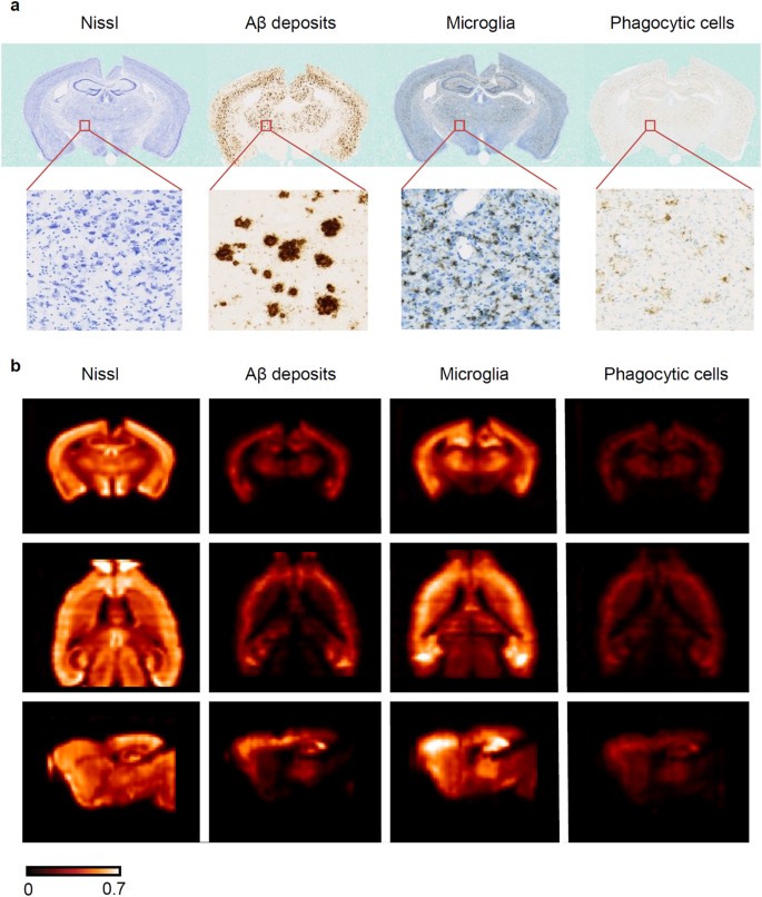
The human proteome in mouse brain - The Human Protein Atlas

Brain Histology and mouse models 2018 Spring

2D section images to 3D atlas - Brain atlases - EBRAINS

Optical Histology: A Method to Visualize Microvasculature in Thick Tissue  Sections of Mouse Brain | PLOS ONE
Optical Histology: A Method to Visualize Microvasculature in Thick Tissue Sections of Mouse Brain | PLOS ONE
Frontiers | The neuroterrain 3D mouse brain atlas

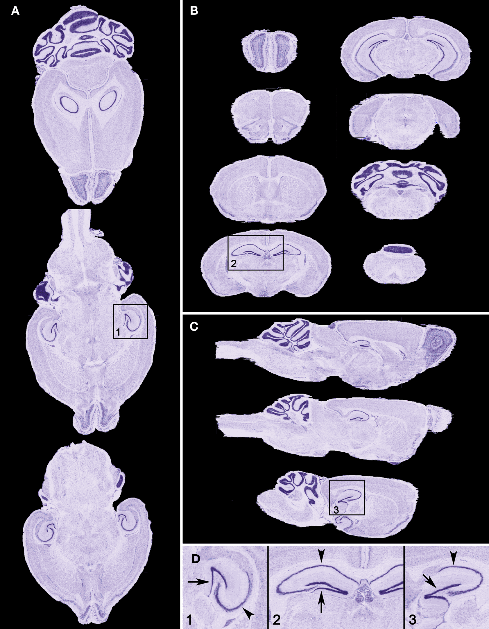
Atlas Thumbnails :: Allen Brain Atlas: Mouse Brain

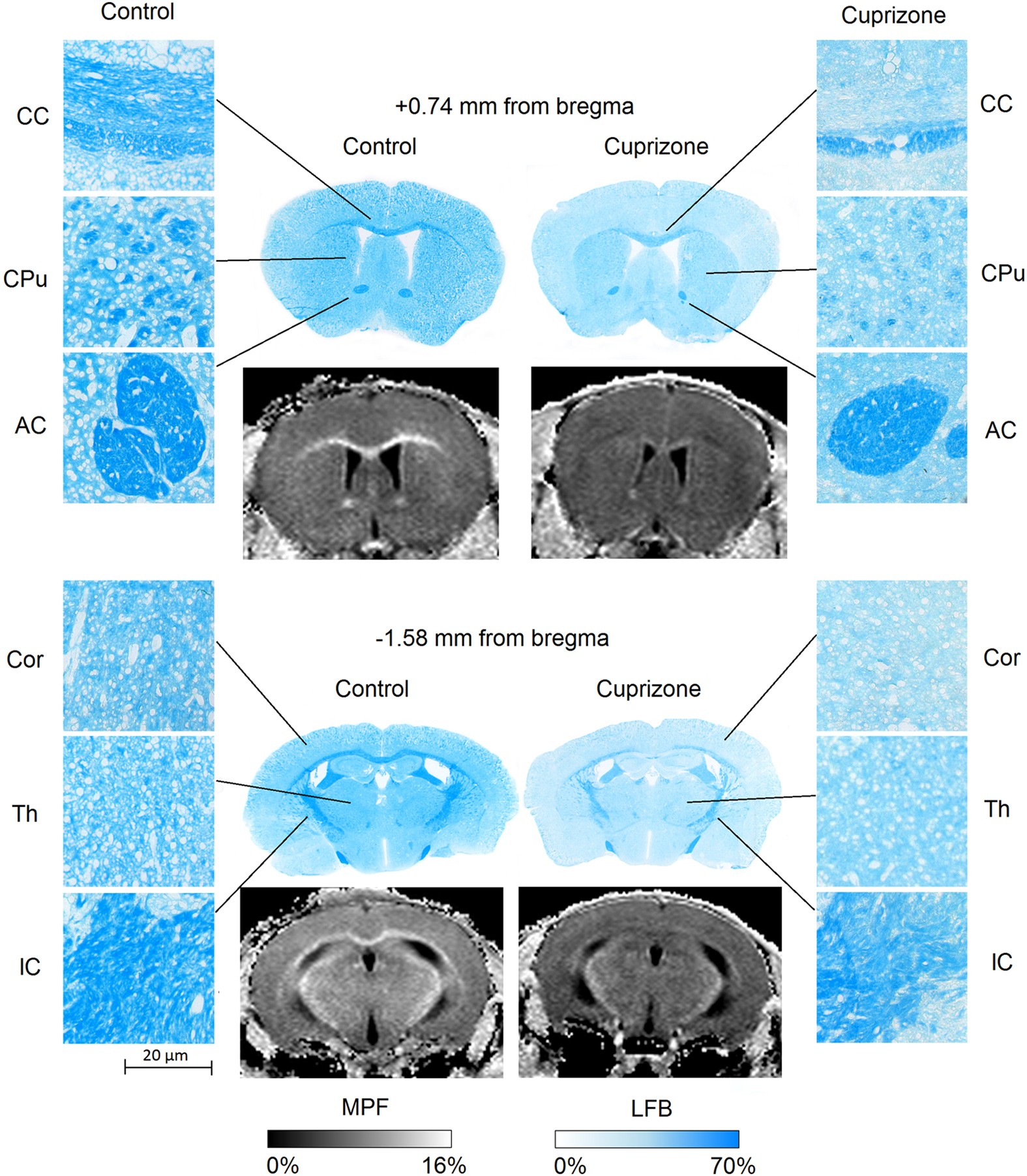
Histological validation of fast macromolecular proton fraction mapping as a  quantitative myelin imaging method in the cuprizone demyelination model |  Scientific Reports
Histological validation of fast macromolecular proton fraction mapping as a quantitative myelin imaging method in the cuprizone demyelination model | Scientific Reports

3D-Registration tool for H&E mouse brain coronal sections - Image Analysis  - Image.sc Forum
3D-Registration tool for H&E mouse brain coronal sections - Image Analysis - Image.sc Forum

Brain Sciences | Free Full-Text | Hippocampal and Cerebellar Changes in  Acute Restraint Stress and the Impact of Pretreatment with Ceftriaxone
Brain Sciences | Free Full-Text | Hippocampal and Cerebellar Changes in Acute Restraint Stress and the Impact of Pretreatment with Ceftriaxone
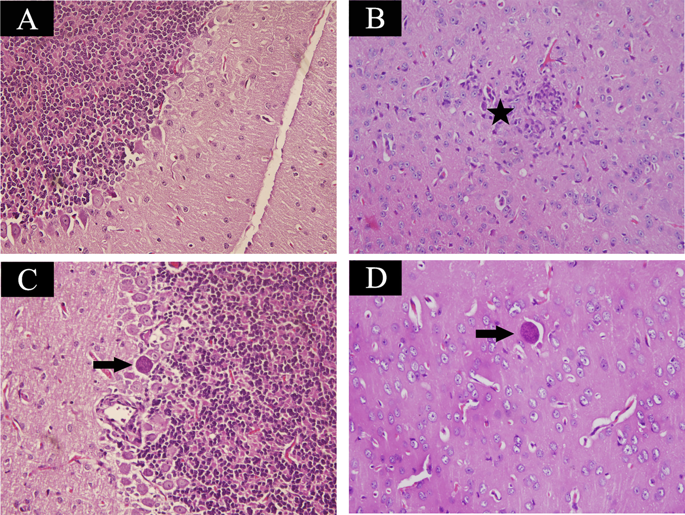
Frontiers | Quantitative Peptidomics of Mouse Brain After Infection With  Cyst-Forming Toxoplasma gondii
Frontiers | Quantitative Peptidomics of Mouse Brain After Infection With Cyst-Forming Toxoplasma gondii

Figure S1. Histology of the adult mouse brain analysed by HE staining.... |  Download Scientific Diagram
Figure S1. Histology of the adult mouse brain analysed by HE staining.... | Download Scientific Diagram

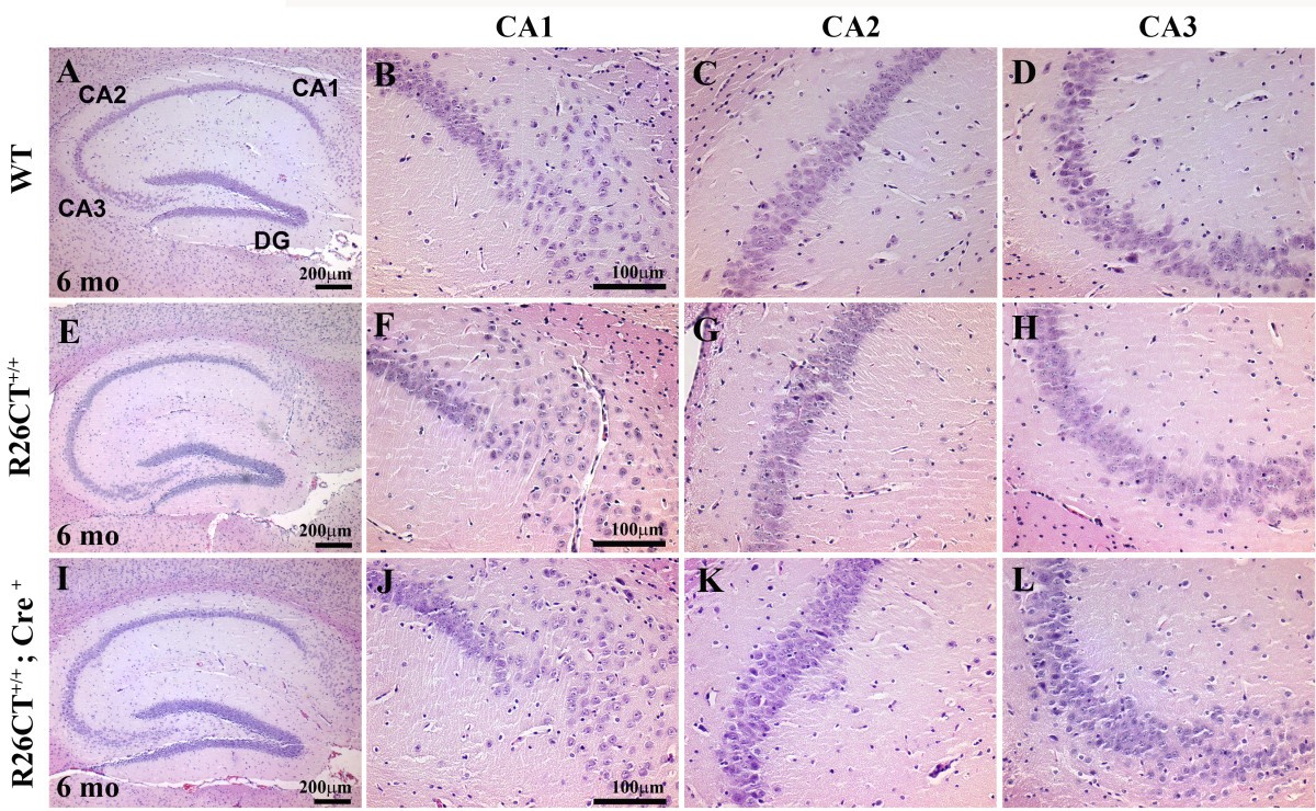
Transgenic mouse model for the formation of Hirano bodies | BMC  Neuroscience | Full Text
Transgenic mouse model for the formation of Hirano bodies | BMC Neuroscience | Full Text

A diffusion MRI-based spatiotemporal continuum of the embryonic mouse brain  for probing gene–neuroanatomy connections | PNAS
A diffusion MRI-based spatiotemporal continuum of the embryonic mouse brain for probing gene–neuroanatomy connections | PNAS

Complete Correction of Enzymatic Deficiency and Neurochemistry in the  GM1-gangliosidosis Mouse Brain by Neonatal Adeno-associated Virus–mediated  Gene Delivery: Molecular Therapy
Complete Correction of Enzymatic Deficiency and Neurochemistry in the GM1-gangliosidosis Mouse Brain by Neonatal Adeno-associated Virus–mediated Gene Delivery: Molecular Therapy

Histopathological examination of the brain of normal mice which... |  Download Scientific Diagram
Histopathological examination of the brain of normal mice which... | Download Scientific Diagram

Histological analysis of brain in Reln-del mice. (a-c)... | Download  Scientific Diagram
Histological analysis of brain in Reln-del mice. (a-c)... | Download Scientific Diagram

Brain Histology and mouse models 2018 Spring

High Resolution Mouse Brain Atlas - Methods

JCI - VEGF antagonism reduces edema formation and tissue damage after  ischemia/reperfusion injury in the mouse brain
JCI - VEGF antagonism reduces edema formation and tissue damage after ischemia/reperfusion injury in the mouse brain

High-throughput 3D whole-brain quantitative histopathology in rodents |  Scientific Reports
High-throughput 3D whole-brain quantitative histopathology in rodents | Scientific Reports

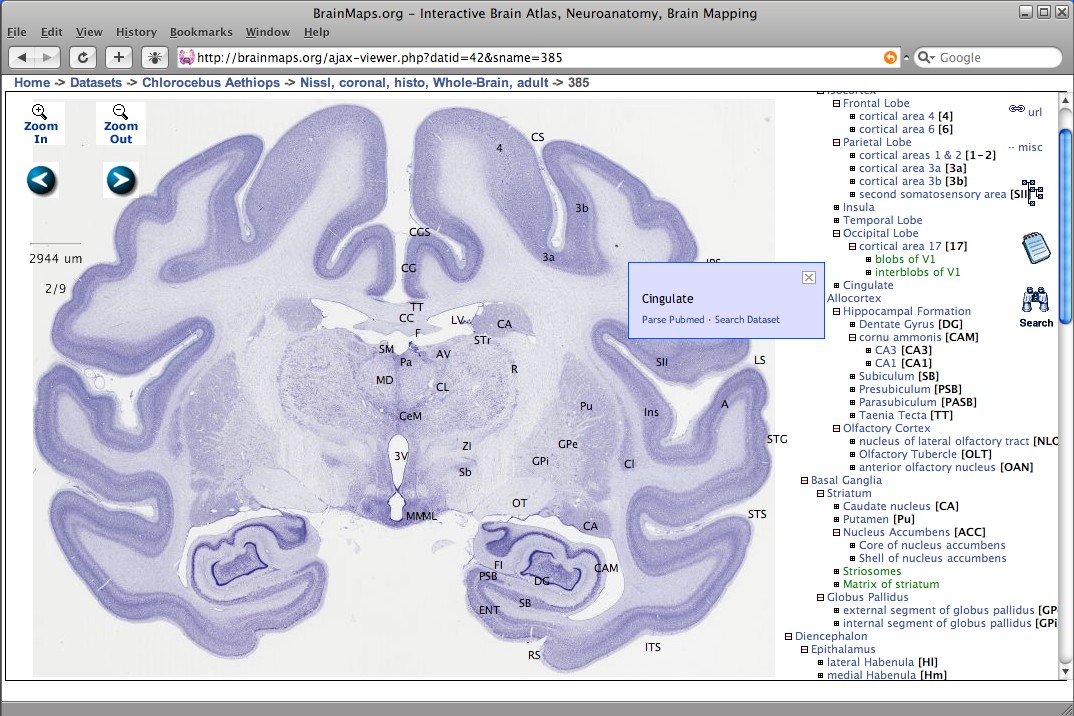
BrainMaps - Wikipedia

Dissection of the long-range projections of specific neurons at the  synaptic level in the whole mouse brain | PNAS
Dissection of the long-range projections of specific neurons at the synaptic level in the whole mouse brain | PNAS

Cytoarchitecture of the mouse brain by high resolution diffusion magnetic  resonance imaging - ScienceDirect
Cytoarchitecture of the mouse brain by high resolution diffusion magnetic resonance imaging - ScienceDirect
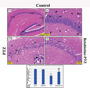
Betahistine Attenuates Seizures, Neurodegeneration, Apoptosis, and Gliosis  in the Cerebral Cortex and Hippocampus in a Mouse Model of Epilepsy: A  Histological, Immunohistochemical, and Biochemical Study | Microscopy and  Microanalysis | Cambridge Core
Betahistine Attenuates Seizures, Neurodegeneration, Apoptosis, and Gliosis in the Cerebral Cortex and Hippocampus in a Mouse Model of Epilepsy: A Histological, Immunohistochemical, and Biochemical Study | Microscopy and Microanalysis | Cambridge Core

Histological structure of mouse brain infected with S. mansoni on day... |  Download Scientific Diagram
Histological structure of mouse brain infected with S. mansoni on day... | Download Scientific Diagram